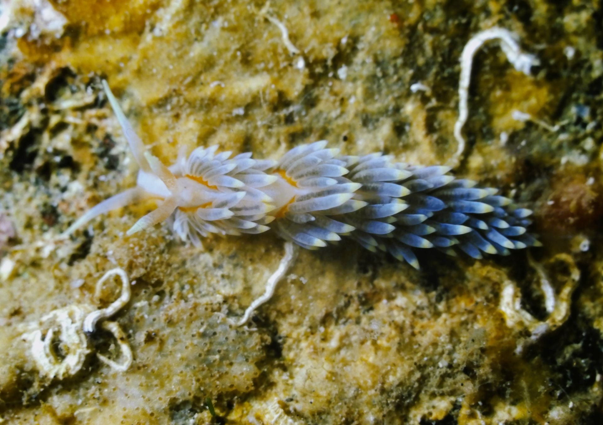
Cover image: Berghia stephanieae. Point of Fort Jeudy, Grenada. 14 feet deep, 24 August 1986. Photo by Hans Bertsch. Reprinted with permission from The Slug Site.
Article by Nora Lowe
This year’s finals had me feeling especially sluggish, so when I heard that there would be a Biology Seminar on sea slug brains, I thought, ‘how topical!’
Dr. Paul Katz from UMass Amherst gave a talk on Monday, December 11 entitled “Exploring the Brain of the Sea Slug.” He is a Biology professor and a member of the Neuroscience and Behavior Graduate Program with close to 100 publications to his name, making him a leading field expert. He also directs the Initiative on Neurosciences (IONs), through which you can receive a monthly newsblast. Plus, he’s helping to organize the first ever Cephalopod Neuroscience Conference in April 2024 at Woods Hole. At Monday’s seminar, Dr. Katz presented his lab’s work, and I discuss its past, present, and future directions below.
Past Work
Dr. Katz started out with a theme from the tech world that is transferable to biology: “The answers to most ‘why’ questions are history,” in that remnants of past function often dictate current form. He nodded to your iphone keyboard as an example. Ever considered why these letters are arranged in that strange order instead of simply alphabetically? The QWERTY design was intended to keep manual typewriters from jamming, a rationale that is now obsolete but still has a visible legacy for that device in your pocket.
Segueing closer to his own field, he described how human photoreceptors — neurons in your retina that turn light into an electric form your brain can interpret — point away from the pupil. This means light has to pass through blood vessels and lots of different cell types before it even hits your photoreceptors. By contrast, the octopus is lucky enough to have light go directly into its photoreceptors and “all of the circuitry” is located behind it instead. Dr. Katz synthesized this idea by saying, “octopuses do it the right way, and vertebrates do it the bizarre way.” This can be chalked up to history, harkening back to the earlier keyboard scenario.
During development, vertebrates, or animals with a backbone, undergo a process called neurulation, in which the neural tube forms. The neural tube, simply put, is the embryo’s central nervous system; it’s the beginnings of the spine and brain. Meanwhile, octopuses don’t do neurulation, so its photoreceptors form right along the epithelium, which you can think of as skin pointing toward the outside world. “So that’s why their photoreceptors are pointing the right way and ours are backward,” Dr. Katz concluded.
From there, he compared brains of different types of organisms, at which point he displayed a phylogenetic tree, sort of like a family tree, focused on brains. Cephalopods have 108 neurons, and primates like us have 1011. Octopuses therefore are the only other example of a giant brain on this planet. Ours and theirs just happened to evolve independently. According to Dr. Katz, “comparing these two giant brains will provide a basis for determining general principles of brain function,” or nervous system operation.
But this presentation wasn’t about cephalopods — it was on gastropods, whom Dr. Katz affectionately calls “their small brain cousins,” as they have 104 neurons, many orders of magnitudes fewer. A key trait of these gastropods, though, is their large and individually identifiable neurons.
Specifically, Dr. Katz studies nudibranchs, which are called sea slugs in everyday lingo, and are shelless molluscs. He has been studying them for more than two decades because 60 of 3,000 nudibranch species can swim. Next, Dr. Katz introduced the audience to Dendronotus, a nudibranch that moves with left-right body flexions. In other words, it flexes side to side. “It’s as simple a behavior as you can imagine,” Dr. Katz says. “It’s just left-right alternation.”
In 1914, a scientist named T. Graham Brown proposed that a simple neural circuit could be responsible for the alternating movements, which he called a half-center oscillator. Dr. Katz emphasized that “there are literally four cells in this animal’s brain that produce this behavior.” For context, this is only four neurons out of 10,000 total in its brain.
Afterward, we met Melibe, part of Dr. Katz’s “cast of characters” that also swims with a left-right body flexion. The fact this flexion is found in all members of the family that includes Dendronotus and Melibe suggests it is a homologous trait in those species. In Dr. Katz’s words, “this behavior was inherited from the most recent common ancestor of this entire clade.” But the behavior isn’t the only thing that is homologous. The neurons are too. They have a very particular morphology, or structure, in which the neurons have a bended axon, and it is unique to just these few neurons in the entire brain. This is key because we can “go from animal to animal and find this neuron based on these criteria, or we can go from species to species and find one neuron on either side of the brain based on the same criteria.”
This is where things get especially interesting, though. Even though many nudibranch species have this half-center oscillator composed of the very same neurons, they have different connectivity. Dr. Katz went on to say that “different wiring for the same neurons” means “they’re actually functionally different in how they work.” In short: “same cells, same behavior, different neural mechanisms.”
Here is how they know that for sure: a researcher in Katz’s lab chemically blocked the synapses (i.e., where the neurons connect and communicate) of one of the cells in order to remove it from the circuit entirely. In Melibe, this caused the animal to slow down its swimming pattern. It would switch irregularly from right to left. Enter the dynamic clamp, a manipulative technique invented at Brandeis University to model the synapse on a computer, which can subsequently inject current to mimic the synapse. In summary, “we take the synapse out pharmacologically, and we put it back in artificially with the computer.” That’s when something amazing happened: The rhythm was restored. “All we did was reconnect these through the computer. And now it speeds up again.” We are now confident these four cells in the half-center oscillator operate to produce this simple motor pattern.
The researchers took things even further, using the dynamic clamp to rewire Dendronotus’ setup into the Melibe circuit configuration — and it worked! From this, Dr. Katz learned “we can take the circuit topology from one species and impose it on the brain of another species, causing it to behave like the other species.”
Current Work
The only problem is to study these animals, you need to ship them over from the Pacific Ocean, which is very costly. In addition, weather or supply chain problems result in a shortage of animals to work with.
When Dr. Katz was at Georgia State, an undergraduate in his lab made major strides by establishing that a sea slug species called Berghia stephanieae can be raised in the lab and mature from egg to adult in less than two months. Over just six weeks, the number of neurons in its brain increases rapidly. Important to note is that humans do the opposite and lose neurons as we age.

Some team members in the Katz Lab study sea slug behavior. They have observed how Berghia doesn’t swim. It eats anemones and has horn-like cerata that can repurpose the anemone’s stinging nematocysts, toxin-containing cells, as a chemical defense.
A Katz Lab postdoc is studying Berghia mating rituals. It has only 5,000 neurons in its brain yet demonstrates complex behaviors. When two slugs meet, they move their tentacles aside and “kiss.” Berghia are hermaphrodites, too, meaning they’re both male and female simultaneously. After they exchange sperm, they actually cuddle for six hours!
The cuddle is for more than romance. “They’re preventing the other one from cheating,” explained Dr. Katz. “There’s this implied agreement between hermaphrodites: ‘I’ll give you sperm, you raise my eggs, you give me sperm, I’ll raise your eggs. If I eat sperm, I broke the contract.’”
One way to circumvent this behavior sequence is with egg laying hormone identified by a Katz team member. That same peptide, or protein, hormone is found in lots of different molluscs. It can be synthesized and artificially injected into Berghia to produce eggs. Oddly enough, they found egg laying hormone expressed in peripheral neurons. “We don’t know what this is for yet,” mentioned Dr. Katz, “but it’s very interesting that they have this peptide hormone throughout the brain and throughout the periphery.”
Still, other recorded behaviors have to do with visual perception. A Katz Lab PhD student found that if you put a stripe in their tank, they straighten out their paths and use their eyes to navigate even though they only have four photoreceptors.
Dr. Katz lamented that even though Berghia, just as other nudibranchs, has easily identifiable neurons, lots of its genes are unannotated. This means we don’t know what they are because no one’s ever studied them before. Dr. Katz attributes this gap in knowledge to the popularity of other model organisms like Drosophila (fruit flies), mice, and zebrafish, who sometimes overshadow nudibranchs in the research world.
Nevertheless, he maintains that nudibranchs offer unique knowledge to neuroscience. For example, serotonergic neurons that express tryptophan hydroxylase, an enzyme precursor to serotonin (the famed chemical messenger) also express unc-4, which is a transcription factor that has been found in C. elegans (i.e., roundworms) and Drosophila. Normally, unc-4 is found in motor neurons expressing acetylcholine, a different neurotransmitter than serotonin. “If you only studied Drosophila and C. elegans, you would think unc-4 determines cholinergic neuron type, but it doesn’t. Instead, the neurotransmitters are a secondary property that comes along.”
Future Work
The Katz Lab is also collaborating with a Harvard lab to do electron microscopy. This involves cutting the nudibranch brain into thousands of sections and putting it onto tape fed through an electron microscope. The goal was to see all the synapses “in one fell swoop,” but COVID interrupted this effort. Chance would have it, however, that this hurdle led to a new and important line of questioning.
“This is the image that changed my entire view of the nervous system,” Dr. Katz said when introducing a cross section of axons just 100 nanometers in diameter. Their miniscule size puts them “at the theoretical limit of how small axons can be and still conduct action potentials.”
Nevertheless, Dr. Katz’s team applied a machine learning technique originally created for octopus ultrastructure to automatically segment all these teeny tiny nudibranch axons. They counted 30,000 total in just that one nerve. These are connected to 9,000 neurons, and the central brain only has 5,000 neurons. This begged the question: “Which is the brain? Is it the part that has a large number of small neurons, or the part that has a small number of large neurons?”
An undergraduate recently also found many boutons, axon enlargements, that are not connected to neurons at all. Surprisingly, they are “engulfed by glial cells,” or nervous system support cells. Dr. Katz stressed, “not only are there giant neurons, but also there are giant glial cells that are as big and encompassing as neuronal cells.”
On top of that, evidence indicates the whole Berghia body is filled with neurons. They’re not just in the brain, but also in the epithelium. Dr. Katz said one of his undergrads even found 12 different neuropeptides scattered all throughout nudibranch physical structures. “The nervous system extends throughout the head. It’s not just in the ganglia,” or nerve cell clusters, Dr. Katz clarified.
This again raises questions about what exactly constitutes the brain of these animals. “Historically, it has been thought that there were particular proliferative zones where neurogenesis occurs,” highlighted Dr. Katz. Nudibranchs are complicating that theory given they have migratory cells, or non-differentiated neurons migrating inward from the skin.
As we learned earlier in the octopus-versus-human photoreceptor case study, vertebrates undergo neurulation in which the central nervous system forms from the neural tube. Neurogenesis occurs in a certain zone, and neurons migrate outward. In mollusks, on the other hand, it seems neurons migrate inward, and there is no neural tube to begin with. “So where do the peripheral neurons come from?” Dr. Katz asked. In short, we don’t know yet.
The Katz Lab is working on understanding the extent of the distinction between brain and body, and he left us with a recap of two more thought-provoking experiments. One of his lab members found photoreceptors in the Berghia eyes projected out to the nose, making it a direct connection between primary sensory neurons. “This is supposed to happen in the brain, not at primary sensory cells,” Dr. Katz recognized.
They were consequently able to connect this finding to prior literature. In 1978, researchers found puffing an odor onto nudibranch photoreceptors caused them to hyperpolarize, or experience a change in charge. Essentially, the photoreceptors were responding to odor stimuli.
Finally, another landmark experiment found that you could cut out the Berghia brain, and the animal can still move around, albeit not as well as when it has its brain intact. “In mollusks, there might not be a clear distinction between central and peripheral nervous systems,” said Dr. Katz. Ultimately, this beautifully unassuming creature actually shines a light on enormous questions about distinguishing body and brain.
You must be logged in to post a comment.